OPERATION
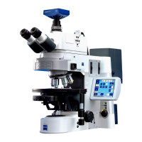
Axio Scope.A1 Illumination and Contrasting Method Carl Zeiss
M60-2-0007 e 05/08 69
4 OPERATION
4.1 Illumination and Contrasting Method
4.1.1 Adjusting the Transmitted Light/Bright-Field According to KÖHLER
(1) General principle of operation
The transmitted light/bright-field microscopy method is among all optical microscopy methods the one
which is most commonly used. High-contrast or tinted samples (e.g. a blood smear) can be examined
easily and quickly.
For an imaging result which is as true to the object as possible we need not only consider the so called
direct bundled beams but just as well the indirect ones, i.e. the beams which diffract and scatter on the
sample details. According to ABBE, the image is more true to the object when the fraction of the indirect
bundled beams is as larger as possible.
The best performance of the microscope, and especially its objective, is achieved when condenser, field
diaphragm and aperture diaphragm are adjusted according to the KÖHLER illumination principle. These
fundamental basic rules for adjusting a microscope are explained in detail in chapter
608H608H4.1.1 (3) "Adjusting
the transmitted light/bright-field according to KÖHLER".
(2) Instrumentation transmitted light/bright-field
Every microscope (except the one with the stand column Vario) is configured to work with the
transmitted light/bright-field method.
All available condensers (except special condensers like dark-field condensers) can be used for the
transmitted light/bright-field method.
(3) Adjusting the transmitted light/bright-field according to KÖHLER
− The Axio Scope.A1 has been put into operation appropriately (chapter
609H609H3).
− The Axio Scope.A1 is turned on.
• Adjust image brightness with light intensity control (
610H610HFig. 4-1/2) on microscope stand.
• Put high-contrast sample into object holder of mechanical stage.
• Bring front lens on condenser (if in use) into place (in objectives ≥ 10x) and turn the gear knob for
vertical adjustment of the condenser (
611H611HFig. 4-1/3 and 612H612HFig. 4-2/2) to the top stop. Make sure the stop is
adjusted in a way to prevent the condenser from lifting out the sample (adjusting the condenser stop,
chapter
613H613H4.1.1 (4)).
• On condensers with revolver/modulator disks: turn the knurled ring (
614H614HFig. 4-2/3) to position H (bright-
field).
• Bring objective 10x in place on nosepiece (
615H615HFig. 4-1/6) and focus sample with gear knob (616H616HFig. 4-1/1).
• Close field diaphragm (
617H617HFig. 4-1/5) enough to make it visible in field of view (even if blurred)
(
618H618HFig. 4-1/A).
• Lower condenser with gear knob for vertical adjustment until edge of field diaphragm appears sharp
(
619H619HFig. 4-1/B).