EASI ECG Lead Placement 7 ECG, Arrhythmia, ST and QT Monitoring
111
EASI ECG Lead Placement
Using a standard 5-electrode set in EASI lead placement you can monitor up to 12 standard ECG leads
simultaneously and continuously at the bedside. EASI provides a monitoring method for trending ST
segment changes that can provide an early indication of ischemia.
WARNING EASI-derived 12-lead ECGs and their measurements are approximations to conventional 12-lead
ECGs. As the 12-lead ECG derived with EASI is not exactly identical to the 12-lead conventional
ECG obtained from an electrocardiograph, it should not be used for diagnostic interpretations.
Respiratory monitoring is also possible with the EASI placement; respiration is measured between the I
and A electrodes.
Place the electrodes as accurately as possible to obtain the best quality EASI measurements.
When EASI lead placement is selected, EASI is shown beside the 1mV calibration bar on the ECG
wave on the display, and EASI is marked on any recorder strips and printouts.
EASI Monitoring During INOP Conditions If one of the derived EASI leads has an INOP
condition (for example, LEAD OFF), a flat line is displayed. After 10 seconds, the directly acquired
EASI AI, AS, or ES lead (depending on which is available) is displayed with the corresponding lead
label. This causes an arrhythmia relearn.
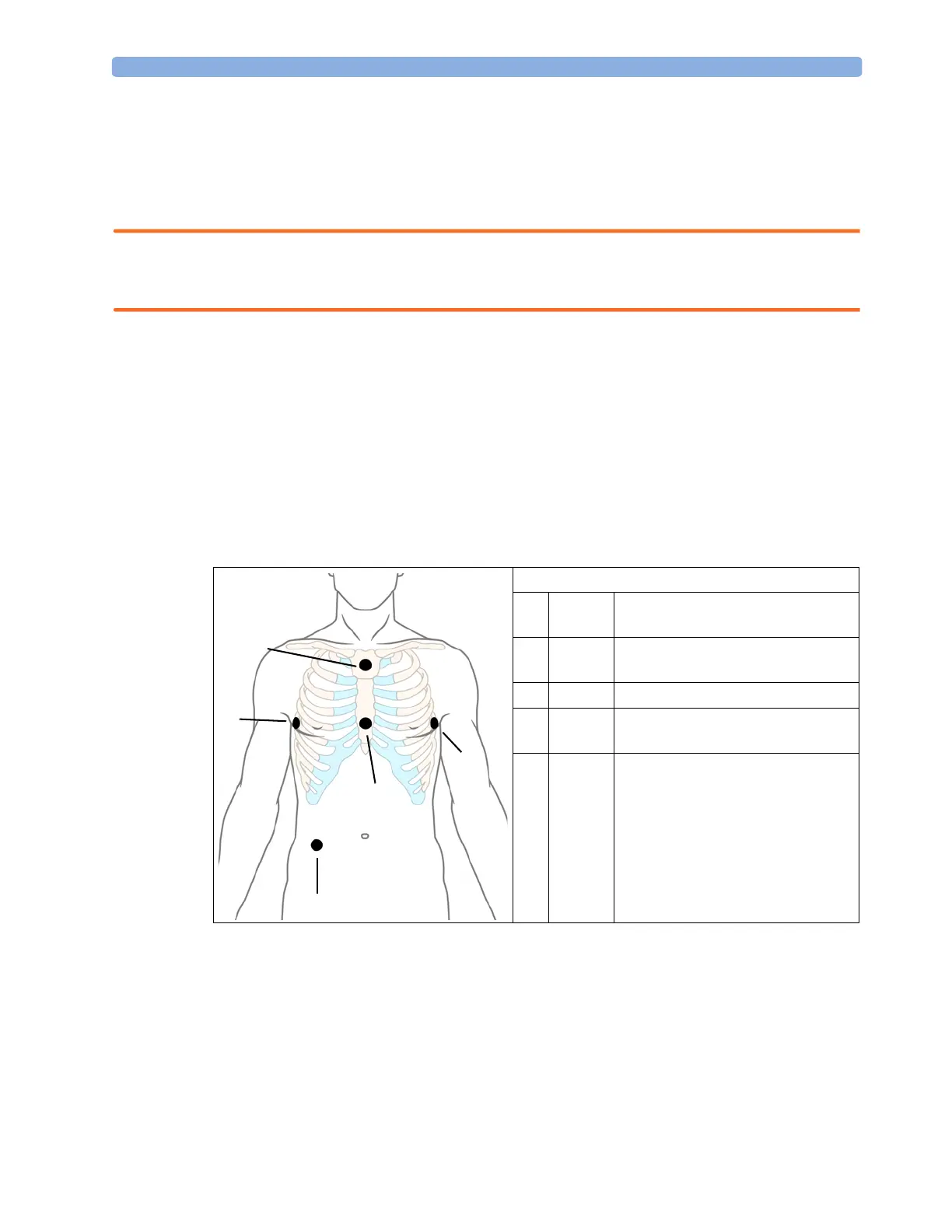
EASI Electrode Placement
1 E (V)
on the lower sternum at the level of
the fifth intercostal space
2 A (LL)
on the left midaxillary line at the
same level as the E electrode
3 S (LA)
on the upper sternum
4 I (RA)
on the right midaxillary line at the
same level as the E electrode
5 N
reference electrode - can be anywhere,
usually below the sixth rib on the
right hip
1
2
3
4
5
 Loading...