Part B - System description cobas p 512
2-18 Operator's Manual - Version 1.6 - 10/2015
NOTICE
To toggle to live image press keys in short sequence
[Fn] + 2x[Scroll] (black keyboard)
[Ctrl.] – [Ctrl.] – [Enter] (white keyboard)
CAUTION
In the screenshots shown below, use the button found only on the keyboard and
stick! Do not use the touch screen. This will activate the functions of the
overlaid screen mask in the QNX control computer and cause a change to the
current operating mode.
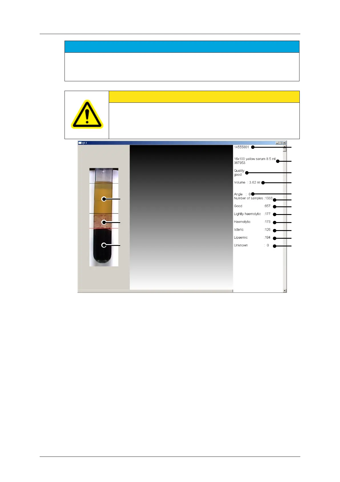
Figure 2-13: Example image (as seen on the monitor)
Turning angle until serum is no longer
occluded by the barcode label
Total number of processed sample tubes
number of "good" samples *
number of "light hemolytic " samples *
Barcode of the current sample tube
number of "hemolytic " samples *
Type of the current sample tube
number of "icteric" samples *
Serum quality of the current sample tube
number of "lipaemic" samples *
Liquid volume of the current sample
number of samples of unknown quality *
* out of the total amount (J)
The image is scanned line-by-line, and the color borders are used to distinguish the
individual phases. The serum is recorded with the color values and the volume is calculated.
The image of the tube is stored in the QS I computer. A results log is sent to the QNX
computer for further processing.
A
 Loading...