Do you have a question about the Siemens ACUSON Cypress and is the answer not in the manual?
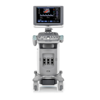
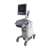
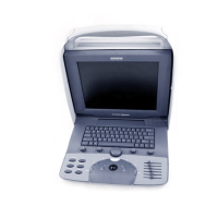
Provides a visual diagram and labels of the main components of the Cypress System.
Detailed steps on how to power on and power off the Cypress system, including warnings.
Explains the process of logging into and out of the Cypress system for user account management.
Guides on creating, managing, and configuring user accounts and their privileges on the system.
Provides instructions on how to properly connect and handle ultrasound transducers with the system.
Details the functionality and usage of the MO disk drive for data storage.
Illustrates and describes the various external ports and their connectivity options on the system.
Provides precautions and methods for safely moving and transporting the Cypress system.
Explains the optimal monitor tilt and viewing angle for clear image display.
Describes the security features available to protect the system against theft.
Details the placement of the system's fan and vents to ensure proper airflow.
Presents a diagram of the Cypress system keyboard and its main components.
Describes the function and operation of each mode key (2D, M, Color, CW, PW, Freeze, Q SAVE).
Explains the use of the trackball for cursor control and <ENTER> keys for input.
Details the functions of the Main knob across different scan modes and controls.
Describes how the soft window knobs control functions displayed at the bottom of the screen.
Explains the purpose and operation of the six dedicated function keys on the keyboard.
Provides instructions on using alphanumeric keys for text input and special character entry.
Details how to create lowercase and uppercase characters with diacritical marks.
Introduces the Setup menu and how to navigate its options and fields.
Explains how to browse and select options within the Setup menu using '+' and '-' signs.
Describes the User Accounts feature and its relation to system security and logon.
Details the various fields available within the Setup menu and their functions.
Explains the Presets feature for storing and recalling system settings for transducers.
Provides steps to save custom system configurations as user-defined presets.
Guides on how to load previously stored user-defined or factory presets.
Describes accessing and configuring background system settings.
Explains setup options for using the system's measurement and calculation functions.
Details DICOM setup options for transferring images and data.
Explains how to configure network settings for system identification and communication.
Describes how to manage patient data, studies, and access the patient directory.
Explains the meaning of various icons used to represent patient data storage formats.
Details the functionality of each button within the Patient menu screen.
Describes the three available formats for saving studies: Cypress, DICOM, and Multimedia.
Provides step-by-step instructions for creating, entering, and modifying patient and study information.
Guides on how to register new patients by querying a hospital information system's worklist.
Details the process for initiating a new study for an existing or new patient.
Explains how to modify existing patient and study details within the system.
Describes how to delete studies or entire patient records from the system.
Explains how to view and print patient reports, including text comments.
Provides general information and basic steps for completing a scanning session.
Explains how soft windows and their control knobs are used in various scanning modes.
Details how to access and use the secondary image function control buttons.
Describes the methods for saving scan data as frames, loops, or series.
Explains how to review saved loops using the VIEW function and associated controls.
Describes how to mark or unmark images, loops, and series for specific purposes.
Explains the system data displayed on screen, its variations, and related controls.
Details triggered acquisition modes for ECG-gated scanning techniques.
Describes two methods for adding text labels or annotations to ultrasound images.
Explains the Quick Text method for adding text labels using the system keypad.
Provides steps on how to use the zoom function to enlarge portions of a scanned image.
Details the 2D sector scanning mode, including activation and controls.
Explains the function of the six slide-pots for adjusting image amplification by depth.
Describes how the Main knob controls Compress and 2D Gain settings in 2D mode.
Details the soft windows and their corresponding knobs used in 2D scanning.
Explains how to activate and use the color flow mapping mode and its controls.
Details the CW Doppler imaging mode, including activation and system controls.
Describes the soft windows and their corresponding knobs for CW scanning.
Explains the function of the TOOLS buttons in CW scanning mode.
Details the PW Doppler imaging mode, including activation and system controls.
Describes the soft windows and their corresponding knobs for PW scanning.
Explains the function of the TOOLS buttons in PW scanning mode.
Details the M-Mode imaging mode, including activation and system controls.
Describes the soft windows and their corresponding knobs for M-Mode scanning.
Explains the function of the TOOLS buttons in M-Mode scanning mode.
Introduces cardiac measurements and categorizes them into general and specific types.
Lists the general measurements available for 2D, M-Mode, and Spectral Doppler.
Lists specific cardiac calculations categorized by 2D, M-Mode, and Doppler modes.
Guides on setting parameters, construction methods, and estimated values for calculations.
Provides steps to make temporary edits to estimated values during a calculation.
Explains how to choose between 'Complete' and 'Configured' report formats.
Describes how to enable and use the averaging function for measurements.
Provides an overview of how to start and use the cardiac calculations package.
Guides on how to perform general cardiac measurements and view results.
Details the process for performing specific cardiac calculations and obtaining results.
Explains how to save measurement results and delete individual measurements from a report.
Provides steps for deleting general measurements from the calculation menu.
Guides on deleting specific measurements from the report menu.
Describes how to correct trace or area outlines using the backspace key.
Explains how to adjust the resolution for trace outlines in measurements.
Details the two methods for accessing and viewing patient reports.
Provides formulas for 2D cardiac measurements and a list of relevant references.
Introduces vascular measurements and categorizes them into general and specific types.
Lists general vascular measurements available for 2D, M-Mode, and Spectral Doppler.
Lists specific vascular calculations for 2D and Doppler modes.
Guides on setting parameters, construction methods, and managing default sites for vascular calculations.
Explains the 'Continuous' and 'Point to Point' methods for defining traces.
Explains how to select the 'Configured' format to display report sections with entered data.
Describes how to add, edit, delete, and manage default vascular sites in calculations.
Provides an overview of how to start and use the vascular calculations package.
Guides on performing general vascular measurements and viewing results.
Details the process for performing specific vascular calculations and obtaining results.
Explains how to save measurement results and delete individual measurements from a report.
Provides steps for deleting general measurements from the calculation menu.
Describes how to correct trace or area outlines using the backspace key.
Explains how to stop a measurement before completion and return to menus.
Provides formulas for 2D vascular measurements and a list of relevant references.
Introduces the Intima-Media Thickness (IMT) measurements package.
Guides on setting up IMT options, including grid and AVI compression.
Lists the two types of IMT calculations: Mean Distance and Standard Deviation.
Provides steps for completing IMT measurements, including trace and site selection.
Details how to connect the ECG signal for display and triggering during stress echo exams.
Explains the importance of ECG signal quality and factors affecting it during stress exams.
Recommends techniques to minimize ECG artifacts for improved signal quality.
Describes the available stress protocols, including stages and view sequences.
Highlights key controls for managing stress exams, such as Protocol, Freeze, and Save.
Explains how to use the PROTOCOL knob to navigate views, labels, and states in stress protocols.
Provides steps to remove unlabeled loops from a patient's study.
Guides on setting up DICOM format for stress studies and image size options.
Describes the TOOLS buttons available in stress echo scanning mode, including the timer.
Provides step-by-step procedures for conducting 2-Stage, 3-Stage, and Pharmacological stress exams.
Explains how the soft windows operate in quad-screen mode for reviewing stress echo data.
Provides steps to view additional loops within the quad-screen display.
Guides on how to remove a label assigned to a loop.
Explains how to reposition the Region of Interest (ROI) box within a quad.
Provides instructions on how to delete loops from a study, noting they cannot be recovered.
Describes methods for saving and labeling loops with stage and view labels.
Explains how to temporarily disable the protocol labeling function.
Details the process of saving a loop and assigning a label to it.
Guides on how to view saved loops in a full-screen or quad-screen format.
Overviews methods for exporting studies and images via MO disk or network.
Details exporting studies and images in DICOM format to an MO disk.
Explains exporting studies in the proprietary Cypress format to an MO disk.
Guides on configuring IP address, subnet mask, and default gateway for network settings.
Details sending DICOM images to a networked DICOM server.
Describes converting images and reports to multimedia formats like AVI and RTF.
Guides on setting up a network connection to send multimedia images to a computer.
Details the DICOM server and Cypress information fields for network configuration.
Describes how to add, edit, remove, and rename server information for DICOM.
Provides steps to specify DICOM settings for network transfers.
Explains how to enable and use DICOM Storage Commitment for verifying image transfers.
Guides on configuring network speed and duplex options for the system.
Details configuring DICOM Modality Worklist settings for patient queries.
Explains how to specify DICOM Storage Commitment settings for server verification.
Provides instructions on physically connecting the Cypress system to a network.
Describes transferring studies to a server, computer, or MO disk.
Explains how to select studies from the Patient List or Study List for transfer.
Guides on configuring transfer settings, destinations, and data for sending studies.
Describes how to remove patient identifying information from transferred images.
Lists common network DICOM transfer errors and their solutions.
Details the operation of the MO drive for transferring studies.
Identifies the parts and functions of the MO disk drive.
Provides methods for formatting disks for use with the Cypress system.
Explains how to read studies stored in Cypress format from an MO disk.
Guides on restoring studies from an MO disk to the Cypress system's hard drive.
Lists common MO disk transfer errors and their solutions.
Highlights critical warnings regarding electrical shock, explosion hazards, and radiation.
Describes the system's intended applications and the types of studies it supports.
Details how cardiac studies obtain images for anatomy and blood flow analysis.
Explains how pediatric studies obtain images for heart and organ abnormalities.
Describes obtaining brain structure images for neonatal cephalic studies.
Explains how peripheral vascular studies obtain images for vessel analysis.
Describes obtaining fetal images for structural and blood flow analysis.
Details how abdominal studies obtain images for organ and vessel analysis.
Addresses hazards that can influence patient safety and methods to reduce exposure.
Provides specific steps to minimize acoustic exposure during scanning.
Highlights warnings related to electrical safety to prevent injury.
Details crucial procedures for infection control, especially for transducer hygiene.
Explains sterilization procedures for noncritical and semicritical device applications.
Discusses biocompatibility of materials used in transducers and latex sensitivity.
Provides information from an FDA medical alert regarding allergic reactions to latex.
Warns about connecting peripheral devices and certification requirements.
Addresses safety concerns for operators, including electrical safety and carpal tunnel syndrome.
Discusses the association of repetitive scanning with CTS and prevention recommendations.
Provides information on OSHA regulations and safe handling of glutaraldehyde.
Details system specifications, classifications, and environmental conditions.
Lists technical specifications including model, manufacturer, power requirements, and classifications.
Provides dimensions and weight of the Cypress system.
Addresses compliance with EMC limits and measures to correct interference.
Explains the meaning of various icons used for system labeling.
Discusses factors influencing measurement accuracy and accuracy by transducer type.
Details the accuracy and range for primary measurements like distance, velocity, time, and heart rate.
Provides accuracy and range for standard derived calculations.
Identifies potential sources of error during scanning and their explanations.
Provides instructions for cleaning the system's external surfaces and display screen.
Details the procedure for cleaning the lens actuator of the MO disk drive.
Covers preventive maintenance practices and transducer inspection guidelines.
Lists basic and regular maintenance practices to prevent system failures.
Provides guidelines for regularly checking transducers for wear or damage.
Lists points to maintain MO disk drive and MO disk performance and reliability.
Advises on managing the internal hard drive for long-term storage of studies.
Provides a guide to the frequency of maintenance procedures for system components.
Outlines basic steps for electrostatic discharge precautions during service.
Provides step-by-step instructions for replacing system fuses.
Details how to clean the trackball and internal surface of the ball-cage.
Provides steps for connecting a video printer to the Cypress system.
Guides on connecting a SVHS VCR to the Cypress system for video recording.
Details connecting both a VCR and a video printer to the Cypress system.
Provides steps for connecting a VGA-compatible monitor to the Cypress system.
Guides on connecting a PS/2-compatible keyboard to the Cypress system.
Provides steps for connecting a compatible ink-jet printer to the Cypress system.
Guides on printing patient reports, including single patient or group reports.
Provides steps to print the patient report of the currently active study.
Explains an alternate method of printing patient reports using the Calculations function.
| Brand | Siemens |
|---|---|
| Model | ACUSON Cypress |
| Category | Medical Equipment |
| Language | English |