110 / 374
SOCT User Manual Version 10.0 rev. A
Fundus photography
• The scan should be overlaid and centered directly on the fovea or optic nerve
head.
• Photo focus should be sharp and clear. Branching blood vessels should be clearly
visible.
• Artifacts that may cast shadows on the OCT scan should be kept to a minimum.
3D Examination Acceptance Criteria
Prior to accepting a 3D examination, the user must ensure that the acceptance criteria are
met.
Saccades
Obstructions in the form of saccades are described in section 8.8.2 Evaluation of OCT
tomograms for Posterior and Anterior scans
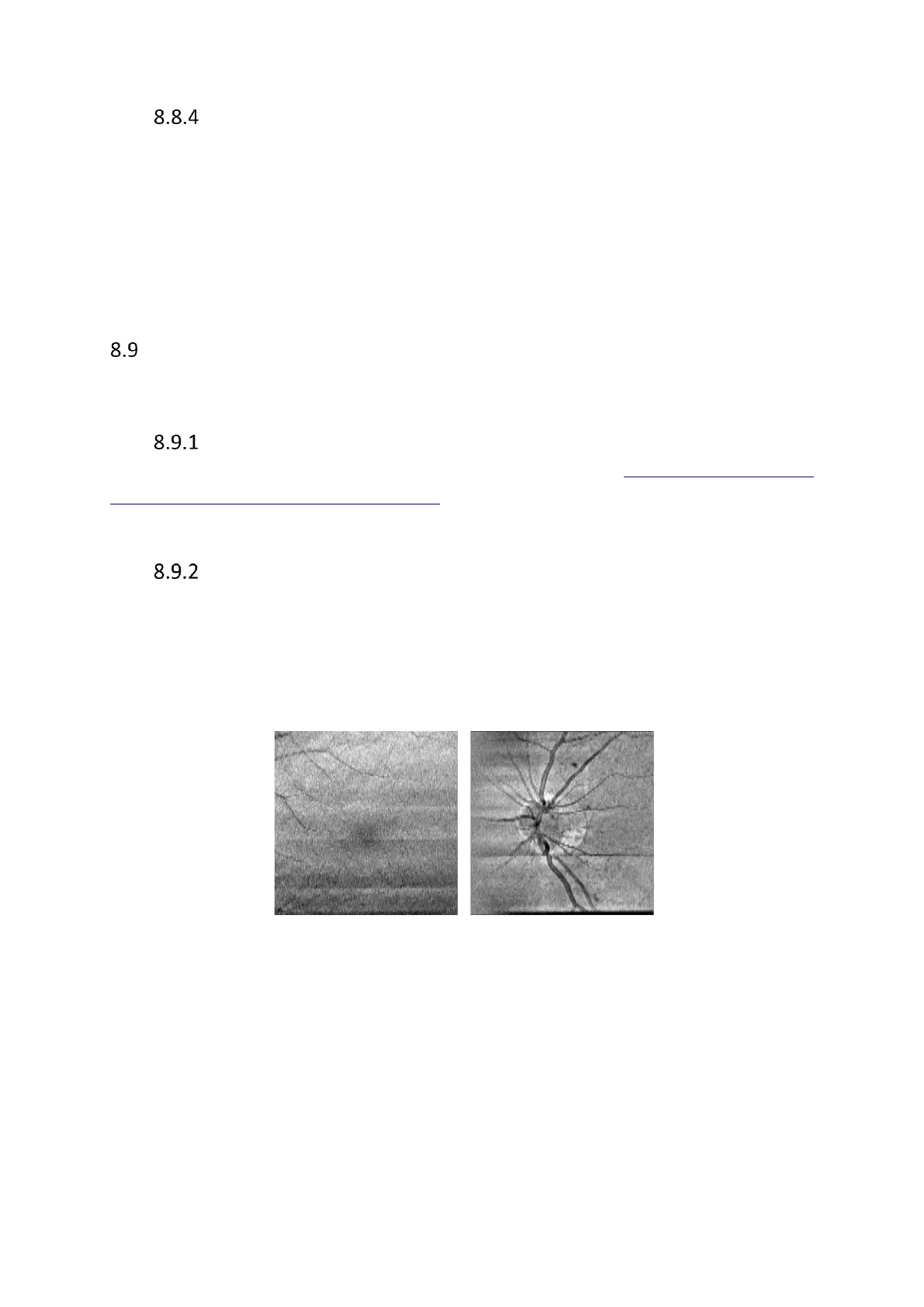
Banding
Carrying out 3D exams with iTracking™ enabled may lead individual B-Scans being acquired at
different horizontal positions. Due to this, there may be vertical tissue variations in the B-scan
window. Although iTracking™ is also purposed with correcting for this motion, it may
nevertheless cause the OCT images to contain intensity artifacts. These artifacts appear as
horizontal lines or form bands in the OCT image, as shown in the examples below:
Given that there are no saccades, exams with OCT images like these should be sufficient for
analysis as there is no protrusion into or through the areas of interest. The operator is
therefore advised to save the exam.