253 / 374
SOCT User Manual Version 10.0 rev. A
microns from the original position of the recognized retinal layer. The negative offset value
describes the position below the original position.
Changing the type of vascular layer on one object affects both eyes and both objects
(angiogram and enface).
NFL thickness map shows the thickness of the NFL layer on the scanned area.
To change the transparency level, turn the mouse wheel over the object.
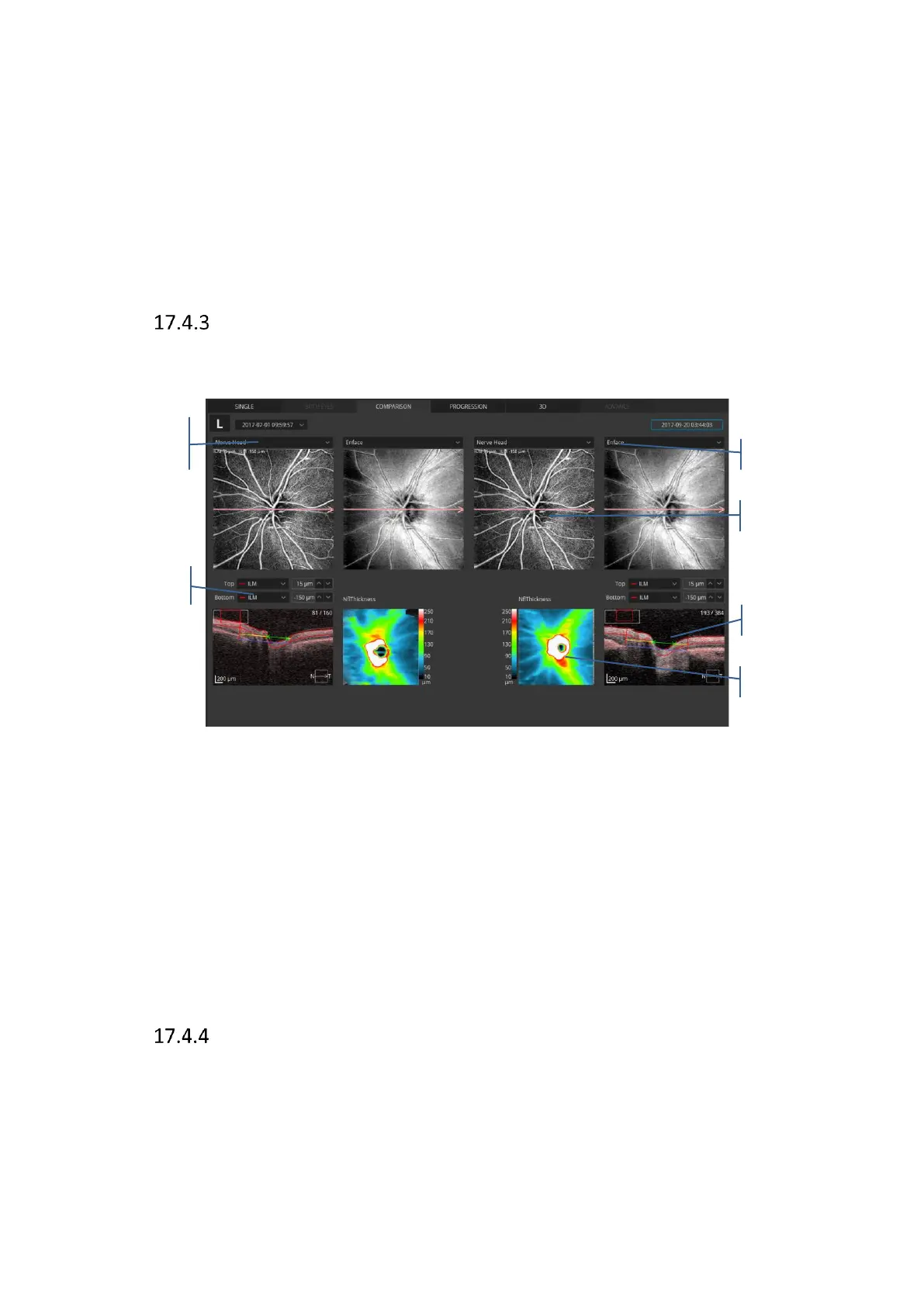
Angio Disc Comparison view
This screen shows the analysis results comparing two examinations of one eye on the same
side in the same scan mode, from different dates.
Figure 254. Comparison Disc Angio view
NFL thickness map shows the thickness of the NFL layer on the scanned area. A map to be
overlaid on the fundus reconstruction can be selected from the list box:
- NFL+GCL+IPL thickness
- GCL+IPL thickness
- NFL thickness
To change the transparency level, turn the mouse wheel over the object.
[Progression] view
This screen shows the analysis results comparing four examinations done on the same side in
the same scan mode, and the same size of scanning area, arranged in a time sequence.
 Loading...


