112 / 374
SOCT User Manual Version 10.0 rev. A
overlying the B-scan. It is important to confirm the presence or absence of Angio flow and
whether it is associated with the layers of interest. It may happen that Angio flow is present in
areas where it shouldn’t be.
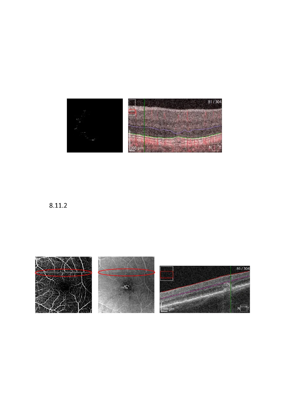
For example, the below image that should be avascular shows several bright areas.
Examination of the B-scan shows an area that has pushed the segmentation up into the hyper-
reflective outer plexiform layer. This was caused by a drusen, therefore any bright signal
detected at this location is likely due to ordinary inner retinal vasculature and should be
regarded as an error.
This example shows an avascular retinal layer which demonstrates that the segmentation is
not correctly passing through the outer retinal layer that is expected to be free of signal.
Image Quality
OCT Angiography is far more sensitive to signal quality than structural OCT imaging. Poor signal
quality will have great effect on image quality and may lead to dark areas, which can affect
interpretation of the exam. OCT Angiography may therefore occasionally display dark spots
that are not a result of capillary dropout but rather due to poor local signal. See examples
below.
It is clear that the issue in this example is caused by saccadic motion. In other cases, floaters
or other media opacities are causes for concern when accepting an OCT Angiography exam.
The operator may also examine the B-scan and the structural enface image.
 Loading...


