86 / 374
SOCT User Manual Version 10.0 rev. A
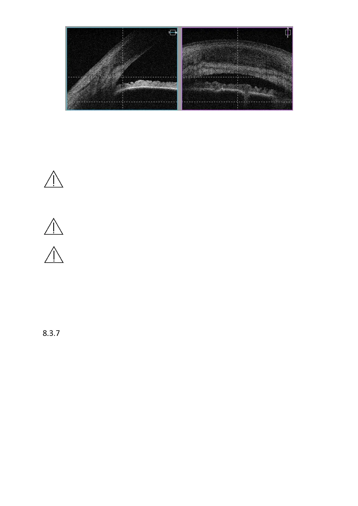
Figure 46.Single angle measurement proper alignment
5) Once the scan location is set in selected place, click twice on the tomogram or press
[Acquire] button. PC will initialize measurement process and then full scan will be
performed.
NOTE: Only when border air - anterior surface is correct are AOD and TISA
measurements accurate. Verify recognition correction before judging the Anterior
Angle morphology.
NOTE: Vertical dense line in the center of Cornea is a natural reflection of laser light
and has no negative influence on measurement result. It can be used to locate the
tomogram in proper position.
NOTE As the Anterior Chamber, wide Angle to Angle and Pachymetry scans are
compensated for beam scanning geometry and reflection from the surface of the
cornea, during acquisition it is important that the scan is centered on the vertex of
the cornea so that a strong vertical reflex is visible through the corneal vertex. The
compensation algorithm works with greatest accuracy when corneal scans are
centered this way.
Wide Anterior programs
8.3.7.1 Wide Anterior programs for REVO FC
The Anterior Chamber Adapter is not applicable for REVO FC models. It has an anterior lens
built in. When a user selects anterior scan program, the lens will automatically install inside
the device.
8.3.7.2 Wide Anterior programs for REVO & NX
The Anterior Chamber Adapter for the SOCT is an easy-to-install hardware attachment to allow
wide scanning of anterior segment structure.
In order to conduct examination of anterior segment, prepare the anterior adapter and follow
the instructions below: