78 / 374
SOCT User Manual Version 10.0 rev. A
Scan optimization and tomogram position alignment. Double click or [Acquire] button starts
measurement.
3. If QI and position of the tomogram are proper proceed to point 6.
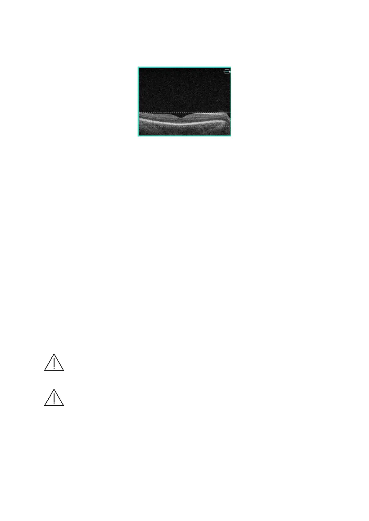
Figure 34. Proper quality and position of the tomogram
4. Manually optimize signal and if required (low saturation, shadows on edges), change the
scanned area e.g. peripheral area.
• Move position of the internal fixation target. Ask patient to follow fixation target,
compensate shadow. The OCT cross-section should be visible in OCT live preview
window. Drag the retina to move tomogram preview to correct position.
• Change scanners offset.
• In order to visualize the interesting retina structures better you can choose
Chorioretinal and Vitreoretinal C-gate mode.
5. Some refracting correction may be needed to obtain the best quality of tomogram.
Observe the QI bar in order to obtain the best signal while changing [FOCUS] bar position.
6. Once the scan location is aligned, ask patient to blink. Click twice on the tomogram or press
[Acquire] button. Device will initialize measurement immediately and then full scan will be
performed.
7. After examination is over the system transfers the captured image into database.
NOTE: If system does not detect the pupil, user has to adjust manually the center
of the patient’s pupil. In order to set working position properly, align the center of
pupil on proper height.
NOTE: In case the system is not able to keep proper position of retina (e.g. patient
is shaking) operator has to switch off tracking and make examination manually.
 Loading...


