61
CAPTURING TOMOGRAMS
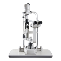
2.7.3. 3D Scan
Move on the inside of the square, which is composed of the given start point and end point, horizontally
and vertically by the step divided by the given resolution.
The scan for [3D Macula] and [3D Disc] is shown below. ("6.0×6.0mm" is fixed for the scan length in both of
these scans.)
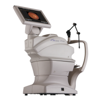
The scan for [3D Macula (V)] and [3D Wide] is shown below. (As the scan length, "7.0×7.0mm" is fixed for
[3D Macula (V)] and "12.0×9.0mm" is fixed for [3D Wide].)
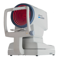
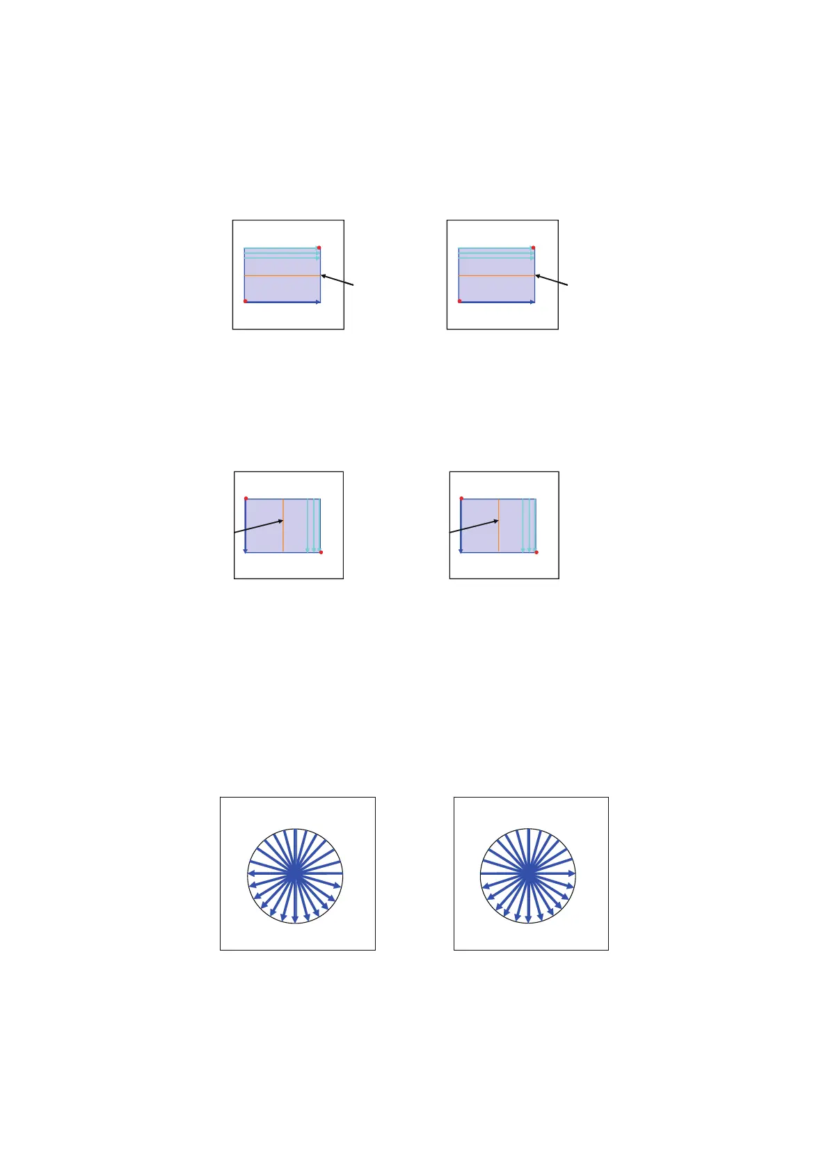
2.7.4. Radial Scan
In the scan range, perform scanning by the specified diameter and by the step divided by the given reso-
lution. "6.0mm" is initially set for the scan length. The start point for Line-Scan and rotating direction are
reversed for each of right and left eyes. For the right eye, rotation is done counterclockwise in the hori-
zontal direction. For the left eye, rotation is done clockwise in the horizontal direction.
[3D-Scan]
End point
Live-Scan
position
Start point
End point
Start point
Live-Scan
position
Left eye
Right eye
S
S
I
I
N
N
T
T
Start point
End point
Live-Scan
position
Start point
End point
[3D-Scan](V)
Left eye
Right eye
S
S
I
I
N
N
T
T
Live-Scan
position
1
2
3
4
5
6
7
8
10
9
11
12
T
S
N
I
N
S
T
I
1
2
4
3
5
6
7
8
11
10
9
12