82
DISPLAYING TOMOGRAMS
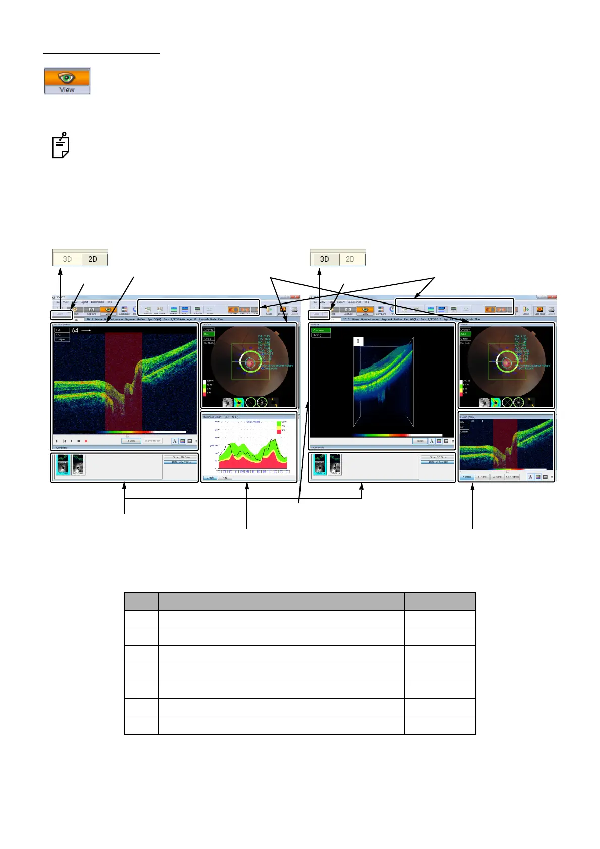
3.4. View Window
Once a tomogram is selected, the View Window is automatically accessed. On View Window,
in 3D/Radial Scan except "3D Macula (V)" and "3D Wide", the 2D/3D tabs are displayed and
you can change the displayed tomogram. In the case of other scans, the tomogram is dis-
played in 2D mode. (The 2D/3D tabs are not displayed.)
See "Drusen Analysis" on P. 162. about the View Window when you use Drusen analysis.
The View Window consists of the following areas.
ID Area 2D/3D
A-1 Thumbnail area 2D/3D
A-2 Image processing controls area 2D/3D
A-3 Fundus/anterior segment image display area 2D/3D
A-4 Thickness graph area 2D
A-5 Tomogram display area 2D
A-5 Auxiliary tomogram display area 3D
A-6 3D tomogram area 3D
2D/3D tabs
2D mode
A-1.
Thumbnail area
A-2.
Image processing controls area
A-3.
Fundus/anterior segment
image display area
A-4.
Thickness graph area
A-5.
Tomogram
display area
A-6.
3D tomogram display area
2D/3D tabs
A-5.
Auxiliary tomogram
display area
3D mode