85
DISPLAYING TOMOGRAMS
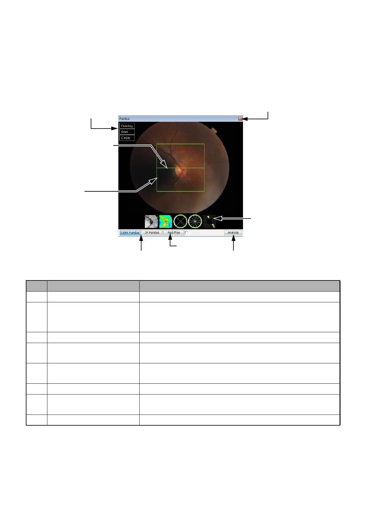
3.4.3. A-3: Fundus/Anterior Segment Image Display Area
Explanation of Window
This window displays the fundus or anterior segment image.
The positional correlation between the image on the fundus/anterior segment image display area and the
tomograms on the 3D/auxiliary tomogram display areas is indicated, including the "overlay" image. For
details, refer to "Pin-point Registration™" on P.121. In this chapter, the fundus image will be explained as
an example.
ID Name Description
C-1 Color and B/W selection Select the color or B/W image. ("Color" is selected as default.)
C-2 Red-free mode Shifts to the red-free image. (When a mark is placed in the check
box at the right of "Red-free", the red-free image is automatically
displayed in the next photography and after.)
C-3 Fundus image analysis Shifts to the fundus image analysis screen.
C-4 Thumbnail overlay image The images which can selected from the "overlay" menu are dis-
played as thumbnails.
C-5 Maximize Maximize this sub-window to the whole screen or return to its
original size.
C-6 Menu Menu (This will be explained in "How to use the menu"on P. 87.)
C-7 Tomogram location indicator The tomogram of the part shown by this indicator is displayed on
Window A.
C-8 3D scanning area This shows the 3D scanning area.
C-1:
Color / B/W selection
C-6:
Menu
C-5:
Maximize
C-4:
Tumbnail overlay
C-8:
3D scanning area
C-7:
Tomogram location indicator
C-3:
Analysis of fundus image
C-2:
Red-free mode