162
ANALYZING TOMOGRAMS
4.8. Drusen Analysis
"Drusen Analysis" helps diagnosis of Age-Related Macular Degeneration (AMD) by detecting and analyz-
ing Drusen whose diameters are 125 µm and over.
Use conditions
"Drusen Analysis" can be used only for the image photographed by the following scan protocol.
When the data is analyzed with "Type2 (New)" of "Segmentation Analysis Type" ("OCTSet Analy-
sis" tab on P.219), the "Drusen" analysis cannot be used for it.
4.8.1. How to use this function
1 Select a patient from the [Search Patient] panel.
2 Select the photography data applicable to the use conditions from the Data list.
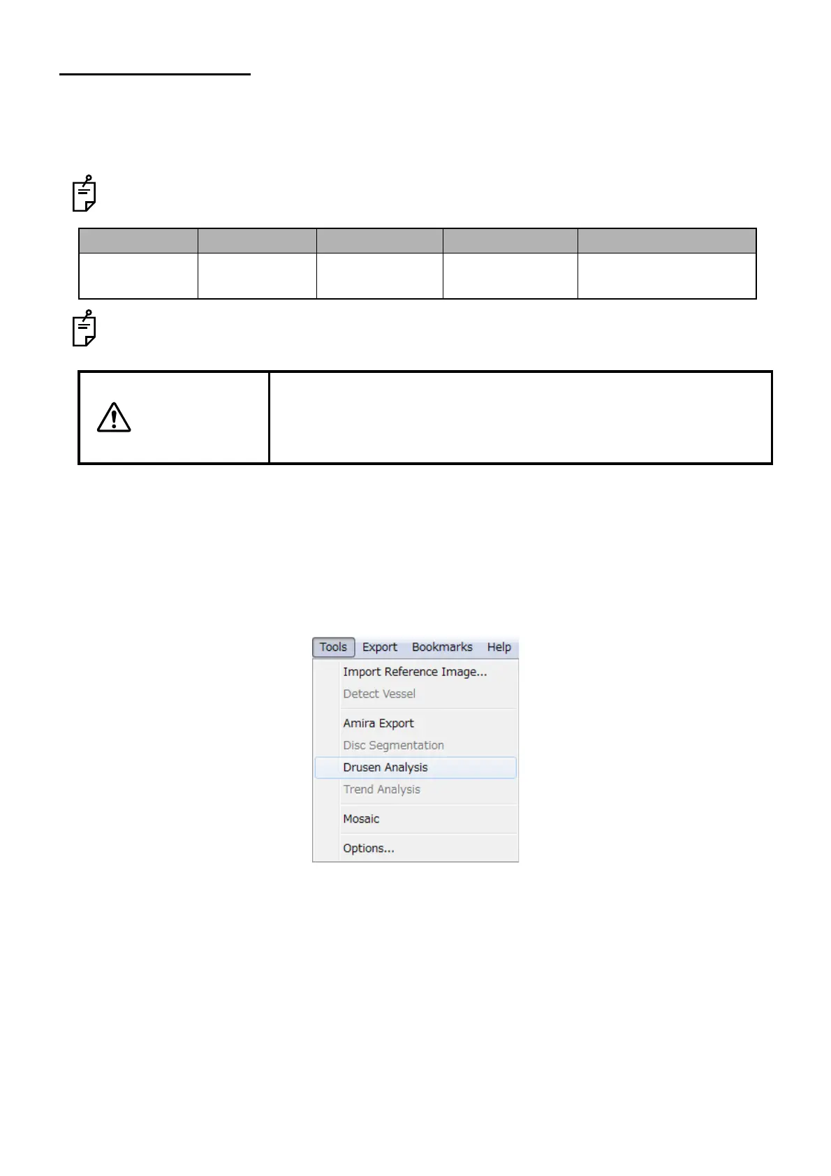
3 Select "Drusen Analysis" from the "Tools" menu in the View Window.
Scan pattern Scan length Scan resolution Fixation Analysis mode
3D 6.0×6.0mm 512×128 Macula and
external fixation
Type 1 - Fine Analysis
CAUTION
When RPE was not detected correctly (If the patients blinked, his/her
fixation moved during scan, the tomogram overlaps the top edge of the
image area, or the image quality is low, this may occur), the analysis
result may not be correct.
 Loading...
