17-19 Operators’ Manual
990400 Rev C 2010 May 1
1-2
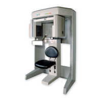
The system consists of a Scanner and Computer Workstation. In
order for the system to operate, both the Scanner and Computer
Workstation must be turned ON.
The system captures data for 3D Skull Reconstruction for the
following procedures:
• Implants
• TM Joints
• Reconstructed Panoramic
• Reconstructed Cephalometrics
• Airway / Sinus, etc.
• Nerve Canal
• PAN - Optional Conventional Digital Panoramic Feature
Cone Beam Volumetric Tomography is a medical imaging
technique that uses X-rays to obtain cross-sectional images of the
head or neck. Quality of the images depends on the level and amount
of X-ray energy delivered to the tissue. Imaging displays both high-
density tissue, such as bone, and soft tissue. When interpreted by a
trained Physician, these images provide useful diagnostic
information.
Intended Use of the Device
The Imaging Sciences International (ISI) Scanner constructs a three
dimensional model from images taken during a rotational X-ray
sequence. The scanner is intended to be used whenever a dentist,
oral surgeon, or other physician needs 3D information of high
contrast objects. The system is designed for imaging of TM joint
studies, mandible and maxilla for implant planning, sinuses, and
other areas of the maxillofacial complex.
U.S. Federal law restricts this device to sale by or on the order of a
dentist or other licensed practitioner.