Do you have a question about the Imaging Sciences i-Cat 17-19 and is the answer not in the manual?
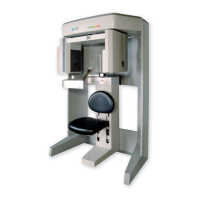
Describes the i-CAT 17-19 Imaging System and its components.
Details the warranty coverage and limitations for the i-CAT system.
Provides guidelines for backing up essential system data.
General safety guidelines for operating the i-CAT system.
General safety principles for radiation exposure.
Overview of the system's built-in safety mechanisms.
Identification and location of various warning and informational labels on the system.
Explains the functions of the operator control box and its indicators.
Details the controls for patient positioning and alignment.
Provides step-by-step instructions for powering on the system.
Guides the user through the proper procedure for powering off the system.
Overview of the patient data management window and study list.
Steps for entering a new patient's details into the system.
Steps for removing a patient record from the database.
Key principles and features for optimal patient positioning.
Using laser alignment lights for precise patient positioning.
Explanation of preview scans and dry runs for positioning verification.
Procedures for performing volume scan acquisitions.
How to create and save custom scan settings for quick access.
Instructions for performing optional panoramic scans.
Overview of the screen displaying reconstructed patient anatomy.
Overview of image filtering options and defaults.
Introduction to the measurement tools available in the software.
Overview of the main screen displaying various image views.
Screen used for planning dental implant procedures.
Tool for marking and estimating the nerve canal location.
Configuration settings for DICOM image handling.
Enabling export of CT and PAN studies.
Procedure for importing patient studies from external sources.
Procedure for calibrating the system's image panels.
Process for calibrating the system's geometric accuracy.
Performing phantom tests to check image quality.
Measuring and evaluating the system's noise levels.
Validating panoramic scan data capture with a phantom.
Overview of preselected scan times and configurations.
Guidelines for safe system operation and installation.
Detailed technical specifications of the i-CAT system.
Operating and storage environmental requirements.
A list of tasks for planned maintenance procedures.
Introduction to network support features for image sharing.
Instructions for installing the Sweeper background service.
Handling situations where network access is unavailable at launch.
Introduction to importing and exporting data remotely.
Instructions for installing and setting up the RSSM module.
How to search for and retrieve images from a remote server.
Minimum hardware and software specs for workstations.
Instructions for installing the iCATVision software.
Overview of tools for manipulating axial slices.
Selecting areas of interest for 3D rendering using Box or Freehand.
Tools for selecting the type of 3D image to create.
Guide to moving around the software interface.
Description of various cursor tools and their functions.
English labels for system components and warnings.
French labels for system components and warnings.
German labels for system components and warnings.
| Imaging Technology | Cone Beam Computed Tomography (CBCT) |
|---|---|
| Scan Volume (FOV) | 17 cm x 19 cm |
| X-ray Tube Voltage | 120 kVp |
| Voxel Size | 0.125 mm - 0.4 mm |
| Software | i-CAT Vision |
| Applications | orthodontics, oral and maxillofacial surgery |
| X-ray Generator | High frequency |
| X-ray Tube Current | 5 mA to 7 mA |
| Power Requirements | 220 V |