17-19 Operators’ Manual
990400 Rev C 2010 May 1
9-2
• Coronal View - double-click displays the MPR Screen.
• Axial View - double-click displays the TMJ Screen.
Detail screens can also be selected from the
Screen menu accessed from the top menu
bar. This enables movement from one detail
screen to another without having to access
the Preview screen.
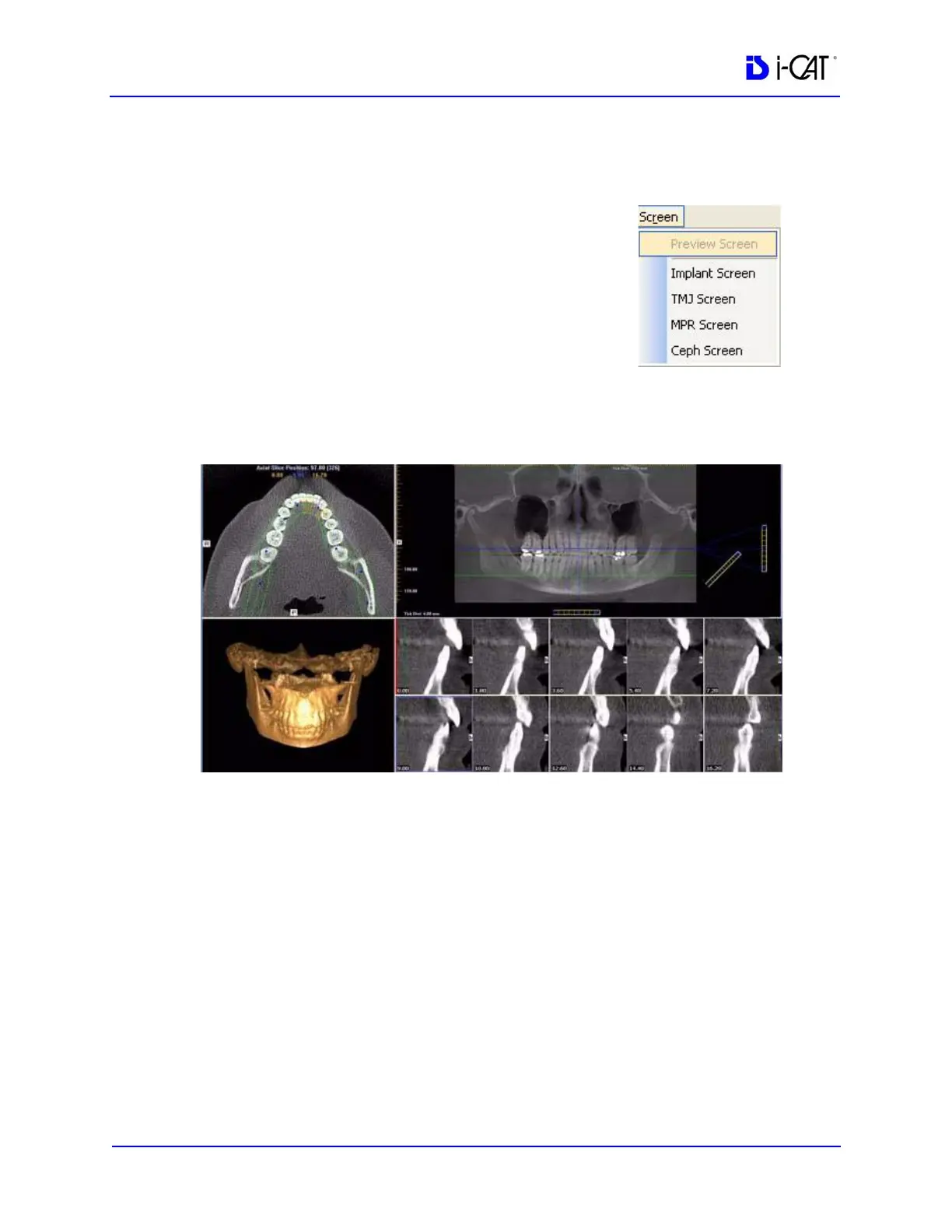
Implant Planning Screen
Double-clicking the Panoramic View on the Preview Screen
displays the Implant Planning Screen.
The Implant Planning Screen is divided into four viewing areas:
• Axial Slice Position View (upper left) used to adjust the
panoramic map view (upper right). The mouse scroll wheel
is active to scroll through the slices.
• Panoramic Map View (upper right) used to modify the
position of the axial views (which are represented on the
axial slice position view) and modify the criteria used to
generate the axial views (lower right).
NOTE: If the study was computed using Tru-Pan, the Panoramic
Map View displays a Tru-Pan label on the image.
• 3D Model View (lower left) shows a three-dimensional
representation of the anatomy of interest displayed on the
Implant Planning Screen. Dragging the cursor across the