4D/Multi-plane LV
Vivid S70 / S60 – User Manual 8-55
BC092760-1EN 01
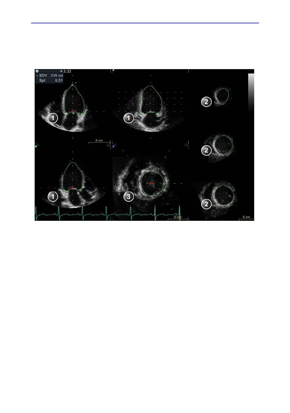
• End diastolic volume
• Sphericity Index
NOTE: You can press Store at any point during the procedure.
Figure 8-28. Left ventricle contour detection (end-diastole)
End-systolic Volume contour detection
1. Select ESV (End-Systolic Volume).
The system automatically displays the loop at the estimated
end systolic frame within the same heart cycle.
2. If the automatic end systolic frame detection is not optimal,
use the Move ES control to set the new end systolic frame.
3. In one of the apical views, place two points: one at the
center of the LV base and one at the apex.
A contour is drawn in all views.
NOTE: If the Auto or Manual contour detection method was used to
define the EDV contour (see step 3 on page 8-54) the same
method is used for the ESV contour detection.
1. Apical views
2. Short axis views
3. Interactive view

 Loading...


