Introduction
User Manual corpuls
3
20 ENG - Version 2.1 – P/N 04130.2
3.3 Description of the Monitoring, Diagnostic and
Therapeutic Functions
3.3.1 Monitoring and Diagnostic Functions
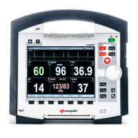
The corpuls
3
has the following monitoring and diagnostic functions:
• ECG
• Diagnostic ECG
• CPR feedback
Optional:
• oximetry (SpO
2,
SpCO
®,
SpHb, SpMet
®
)
• Capnometry (CO
2
)
• Temperature (Temp)
• Non-invasive blood pressure monitoring (NIBP)
• Invasive blood pressure monitoring (IBP)
With the 4-pole ECG monitoring cable, the bipolar extremity leads according to
Einthoven (I, II, III) and the unipolar extremity leads according to Goldberger
(aVR, aVL, aVF) can be derived and displayed on the monitor.
By combining the 4-pole ECG monitoring cable with the complementary 6-pole
ECG diagnostic cable (chest wall leads according to Wilson (C1-C6)), 12
channels can be displayed simultaneously. This enables a comprehensive ECG
diagnosis which can be supported by the ECG measurement HES
®
Light and
an optional ECG analysis software.
During resuscitation, the CPR feedback option monitors the current
compression rate and -depth of the thorax compressions by means of the
corPatch CPR sensor. Speech- and text messages signal to the user whether
the quality of the thorax compressions is sufficient or can to be optimised.
Besides the peripheral pulse rate (PP), oximetry measures the perfusion index
(PI), the arterial oxygen saturation (SpO
2
) in percentage, the level of
methemoglobin (SpMet
®
) and, depending on the used oximetry sensor, the
level of carboxyhemogolobin (SpCO
®)
in percentage or the level of total
hemoglobin (SpHb) in g/dl or mmol/l. Up to six parameter fields with digital
measuring values can be configured for display. A curve field can display the
oximetry plethysmogram.
The capnometer, which works according to the mainstream method, measures
the CO
2
concentration in the patient’s expiratory breath in real time. The CO
2
concentration, measured in mmHg or kPa, can be displayed on the screen as a
capnogram. The corpuls
3
allows use of capnometry in intubated and non-
intubated patients. The patient’s respiratory rate is measured as an additional
parameter.
Up to two temperature values can be measured by means of temperature
sensors and displayed as numerical values: body core temperature rectally
and/or oesophageally and surface temperature.