User Manual corpuls
3
Operation – Therapy
ENG - Version 2.1 – P/N 04130.2 69
The curve field in the first line of the screen is pre-set and cannot be configured.
The first curve field displays the ECG recorded by the respective defibrillation
electrodes, switching automatically between IIauto and DEauto:
• corPatch electrodes: - DEauto recording, via the corPatch electrodes
• shock paddles: - Einthoven lead IIauto,
recorded via ECG electrodes and 4-pole ECG
monitoring cable
or
- DEauto recording,
via shock paddles, if no 4-pole ECG
monitoring cable is connected
The value of ECG amplitude is set to 10 mm/mV. Automatic ECG amplitude
control is disabled.
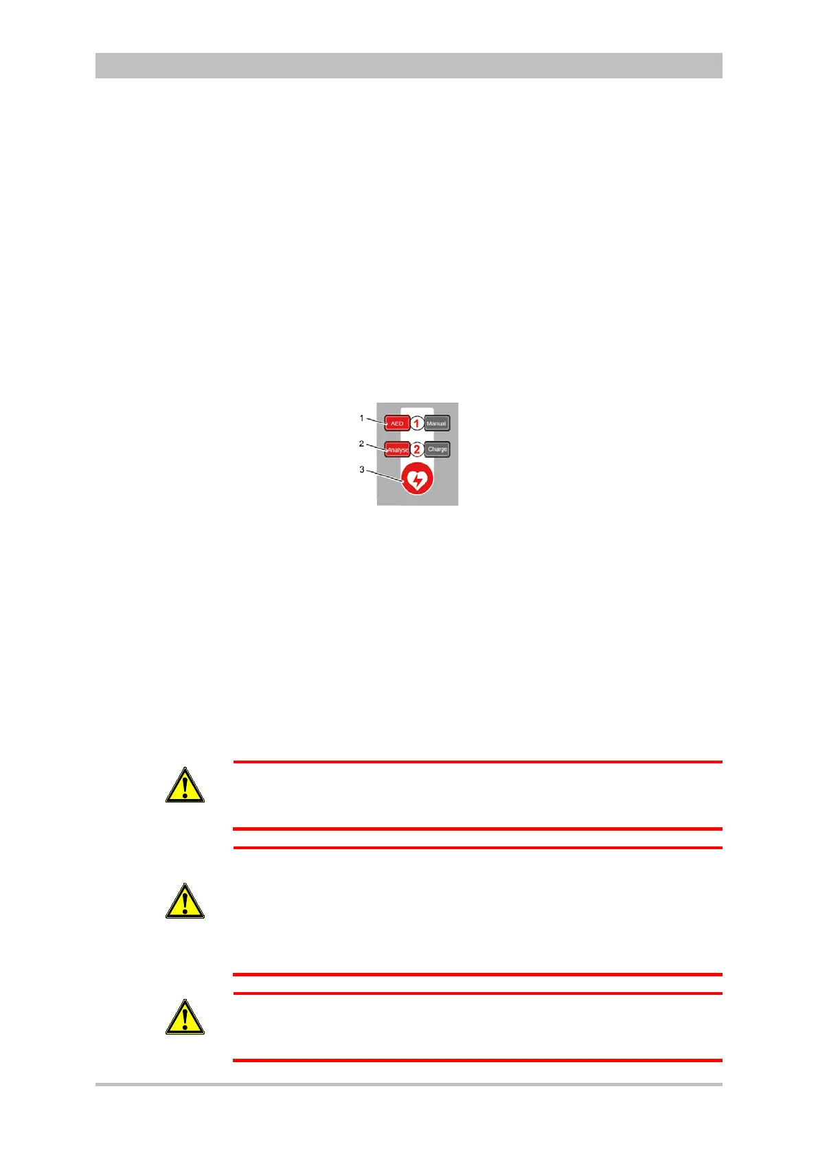
The following keys are available to operate the device in AED mode:
1. AED
2. Analyse
3. Shock
Fig. 5-4 AED mode function keys
When using corPatch electrodes, the shock is released by pressing the Shock
key at the monitoring unit.
When using the shock paddles, the shock is released by pressing both buttons
at the shock paddles.
The charged defibrillator can be discharged internally by pressing the softkey
[Cancel].
In AED mode, a configurable audio recording option is available which is
disabled by default. If the audio recording option is enabled by the person
responsible for the device, all surrounding noises are recorded (see
chapter 7.4.3 Configuration of the Defibrillation Function (Persons Responsible
for the Device) , page 163).
WARNING
In each individual case the trained user determines the progress of treatment
according to medical requirements. The procedure shown here reflects the
operating possibilities of the device.
WARNING
In patients with an implanted pacer placing the therapy electrodes directly
above the pacer unit is contraindicated.
Under certain conditions irreversible damage to the myocardium might be
caused by the invasive pacer electrode.
In such a case, choose the reversed position of the therapy electrodes: below
the left clavicula parasternally and below the right mamilla, approx. 5th
intercostal space at the level of the apex of the heart.
WARNING
In patients with an implanted pacer, it is possible that shockable ECG rhythms
or arrhythmias will only be detected to a limited extent.