NIR images are captured automatically and seamlessly during the scan, from every angle used for the 3D scanning,
and all collected information can then be reviewed using the iTero Element 5D Review tool.
Note: NIR images should be used in conjunction with the current standard of care for caries detection, and do not
replace it.
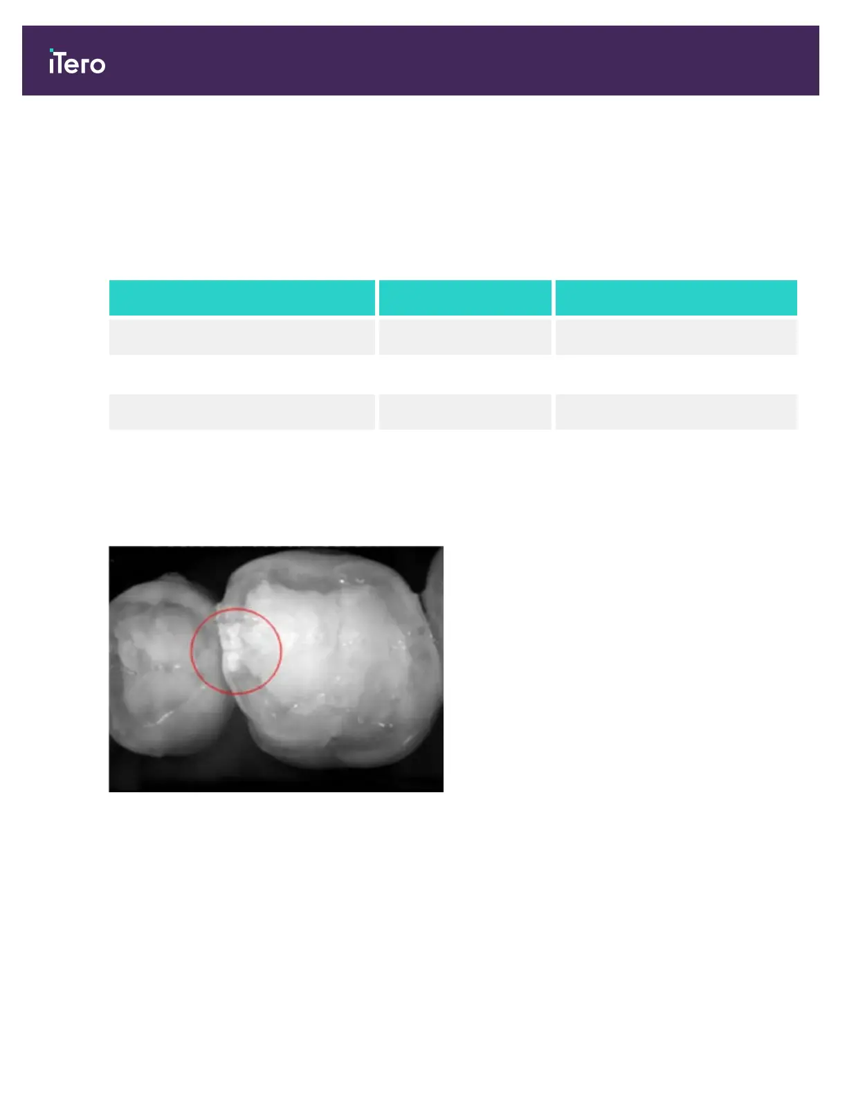
The resulting NIRI grayscale image shows structures with varying translucency as different levels of brightness. The
lower the translucency, the higher the reflection of the infrared light and the brighter the structure. Using this
technology, it is possible to make out the following structures:
Appears Translucency
Enamel Dark High
Interproximal caries Bright Low
Dentin Bright Low
The differentiation between carious lesions and dentin is based upon the location of the bright feature. Dentin is
located in the center of a tooth, whereas interproximal carious lesions appear on the interproximal or distal mesial
region, where healthy enamel is expected.
As such, dentin and interproximal carious lesions appear as bright features, with a dark enamel ring around the dentin
structure, as shown in the figure below, which provides an occlusal view of a carious lesion.
Figure 8: Interproximal carious lesion
iTero Element® 5D Plus User manual
6 © 2020 Align Technology, Inc. All rights reserved.