5‐2MonitoringECGExpressionMR400InstructionsforUse
TheproperplacementoftheECGelectrodesintheMRIiscriticaltoreducingthebloodflow
induceddistortionoftheECGwaveform.WithproperstrategicplacementoftheECGelectrodes
andminimizationofECGleadcablelength,thisbloodflow
induceddistortioncanbekepttoa
minimum,asdiscussedinthissection.Additionalartifactscausedbythestatic,gradientandRF
electromagneticfieldscanseverelydistorttheECG,makingobservationofthemorphologic
changesanddetectionofarrhythmiaquitedifficult.MonitoringusingadifferentECGleadview(I,
II,III,
AVL,AVR,AVF)willminimizesomeoftheseartifacts.
wECG Module and ECG Lead Cable
ThewECGmoduleandleadcableareintendedforpatientuseswhencontinuousECGmonitoring
orcardiacgatingarerequired.ThewECGmoduleandleadcablemaybeusedintheMRsystem
bore,althoughthemodulemustnotbeplacedwithin28cm(11inches)oftheMRIfieldof
view
(FOV).ForwECGmoduledetails,seepage2‐9.ThecomponentsoftheECGleadcablearedetailed
below.
.
• If dropped, the wECG module must be verified for correct operation before use; see page
14-12.
• Guard against the accidental ingress of liquid into the module, as measurements made by the
device can be adversely affected.
Refer to your facility's biohazard procedure for disposal of ECG lead cables when they become
unusable. Usually cables are disposed of as medical waste per facility procedures.
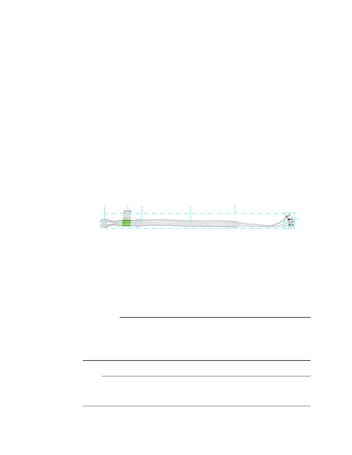
1 Connector
2 ECG lead cable label identifier
3 Velcro storage strap
4 Cable trunk with foam insulator
5 Lead wires
6 Lead cable clips