Optimizing the Image
5-20 LOGIQ 7 Online Help
Direction 2392536-100 Rev. 1
Map
Description The system supplies B, M, and Doppler Mode system maps.
Adjusting To select a map, press Gray Map. A map window displays. The
image reflects the map as you go through the selections.
Values Gray maps gradually change from least contrasty or softest to
most contrasty.
Map values vary by probe, application, and multi frequency
setting. Map values are returned to the factory or user preset
value when you change the following: Probe, Exam Category,
Exam Calcs, New Patient, or Frequency.
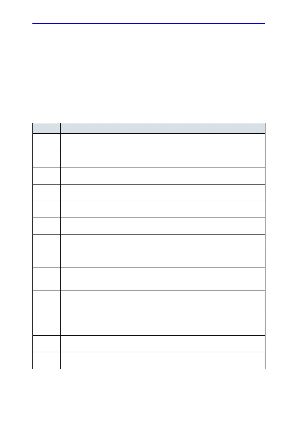
Map Description
A Assigns a greater amount of gray scale pixel values to bright reflectors in the image. Useful
when imaging abdomen, liver, kidney, OB, pelvic, etc.
B Assigns a greater amount of gray scale pixel values to bright reflectors in the image. Useful
when the abdomen, liver, kidney, OB, pelvic, etc.
C Assigns a greater amount of gray scale pixel values to bright reflectors in the image. Useful
when imaging the abdomen, liver, kidney, OB, pelvic, etc.
D Assigns a lesser amount of gray scale pixel values to bright reflectors in the image, compared
to Maps B and C. Useful when imaging bright carotid plaque reflectors.
E Assigns a equal amounts of gray scale pixel values to all reflectors in the image. Useful when
imaging tissue with bright reflectors, e.g., cyst with septations or calcifications.
F Assigns a lesser amount of gray scale pixel values to bright reflectors in the image. Useful
when imaging arteries and grafts.
G Assigns a lesser amount of gray scale pixel values to weak reflectors in the image than Map F.
Useful when imaging small parts.
H Assigns an s-shape to gray scale pixel values. Useful when imaging anatomical signals with
less tissue differentiation, e.g., tendon, vein, carotid, thyroid, breast, etc.
I S-shaped map. This map highlights tissue differentiation for a certain band of signals. Useful
when imaging structures where you want more contrast, e.g., renal, tendon, vein, carotid,
thyroid, breast, etc.
J S-shaped map. This map highlights tissue differentiation for a certain band of signals. Useful
when imaging structures where you want more contrast, e.g., renal, tendon, vein, carotid,
thyroid, breast, etc.
K S-shaped map. This map highlights tissue differentiation for a certain band of signals. Useful
when imaging structures where you want more contrast, e.g., renal, tendon, vein, carotid,
thyroid, breast, etc.
L S-shaped map. This map highlights tissue differentiation for a certain band of signals. Useful
when imaging structures where you want more contrast, e.g., cardiology.
M Assigns a lesser amount of gray scale pixel values to weak reflections in the image than Map
C. Useful when imaging abdomen and liver.
 Loading...