Optimizing the Image
5-82 LOGIQ 7 Online Help
Direction 2392536-100 Rev. 1
Power Doppler Imaging (PDI)
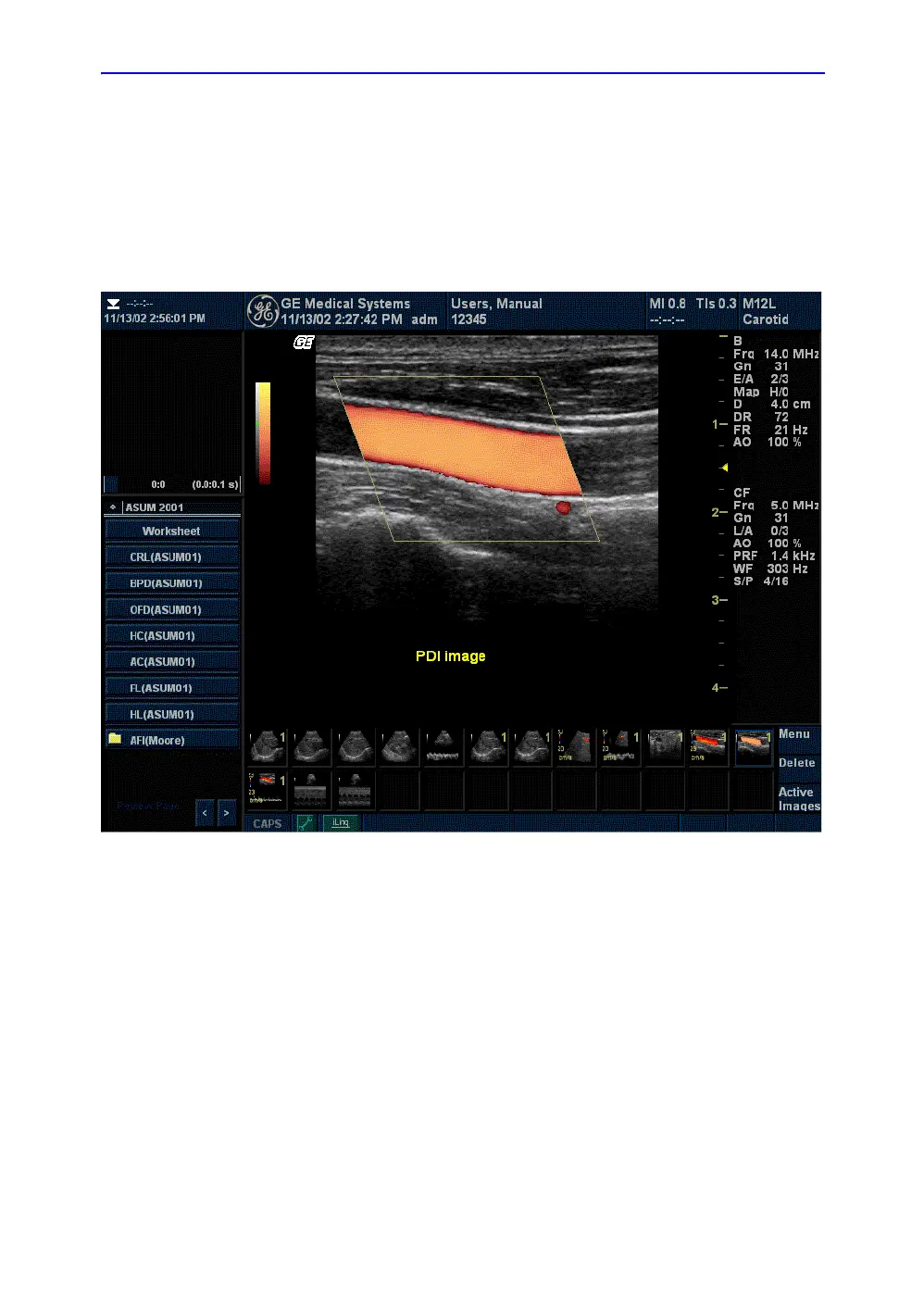
Description Power Doppler Imaging (PDI) is a color flow mapping technique
used to map the strength of the Doppler signal coming from the
tissue rather than the frequency shift of the signal. Using this
technique, the ultrasound system plots color flow based on the
number of reflectors that are moving, regardless of their velocity.
PDI does not map velocity, therefore it is not subject to aliasing.
Figure 5-40. Power Doppler Imaging Display
Adjusting Press PDI. The color flow window appears over the B-Mode
image. Move the Trackball to move the CF window. To exit,
press PDI or select a new mode.
Values On/Off.
Ten power (P0-P6 and P8-P10) and one direction PDI map (P7)
is available.
Benefits Since PDI does not display velocity, it does not aliase.
 Loading...