GE HEALTHCARE
DIRECTION FC091194, REVISION 11 VIVID 7 SERVICE MANUAL
5 - 2 Section 5-2 - General Information
Section 5-2
General Information
5-2-1 Introduction
The Vivid 7 ultrasound scanner can be used with both phased array and linear array ultrasound probes
and Doppler (Pedof) probes.
The system can be used for:
• 2D Gray Scale and 2D Color Flow Imaging
• M-Mode Gray Scale Imaging
• Color M-Mode
• Doppler
• 4D (Realtime 3D, introduced 2004)
• Different combinations of the above modes
Vivid 7 is a digital beamforming system. Signal flow travels from the Probe Connector Panel to the Front
End Electronics, then to the Back-End Processor, and finally, the results are displayed on the monitor
System configuration is stored on the hard drive (inside the Back-End Processor (BEP)) and all
necessary software is loaded from the hard drive on power up.
A Physio module, the Patient I/O, is incorporated in the Back-End Processor (BEP) to provide ECG
signals to synchronize cardiac ultrasound image acquisition. Other analog signals, from devices such
as treadmills (e.g. ECG and phono), are also processed by the Patient I/O.
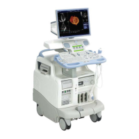
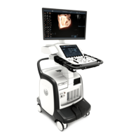
Figure 5-1 Vivid 7 Major Components
NAME PLATE
MONITOR CONTROLS
FRONT COVER
PROBE CONNECTORS
OPERATOR PANEL
B/W PRINTER
BACK-END PROCESSOR CHASSIS
PATIENT I/O
5.25" MO DRIVE (OPTION)
DVD or CD-DRIVE
FRONT BUMPER
PROBE PARKING POSITION
DOPPLER PROBE CONNECTOR
SWIVEL LOCK & BRAKE RELEASE
BRAKE & SWIVEL RELEASE
VCR