EMC
VI-11
Appendix VI Glasgow 12-Lead ECG Analysis
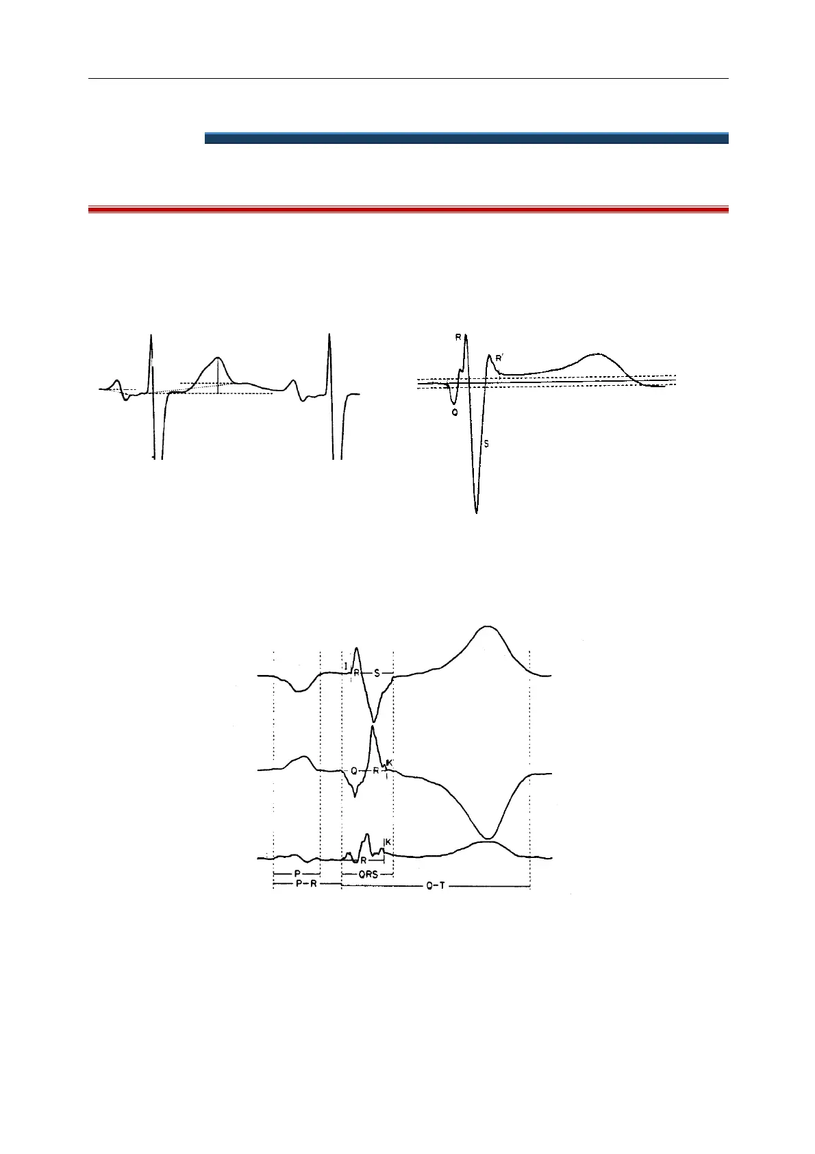
VI.1 Wave measurement
From the 12 average beats, a single combined function is formed and a provisional overall QRS onset and
termination is determined by thresholding techniques. The provisional onset and termination are then used as
starting points for a search for QRS onset and termination within each individual lead.
Figure 1 Varying choice of baselines.
Figure 2 Baseline at the level of QRS onset as used by
the Glasgow program.
In each individual lead, the QRS onset is taken as the baseline and hence Q, R, S, R' waves are measured with respect
to the QRS onset as shown in the accompanying figures from the CSE paper.
Figure 3 Illustration of isoelectric segments I and K.
Isoelectric segments at the beginning of a QRS complex, i.e. a flat segment between the provisional overall onset
and the onset of an individual lead are excluded from the first component (Q or R) of the QRS complex as
recommended by the CSE group.Similar considerations apply at the end of the QRS complex (see Figure 3).