FUNCTIONAL DESCRIPTIONS
Sysmex SF-3000 Operator's Manual -- Revised September 1995 10-3
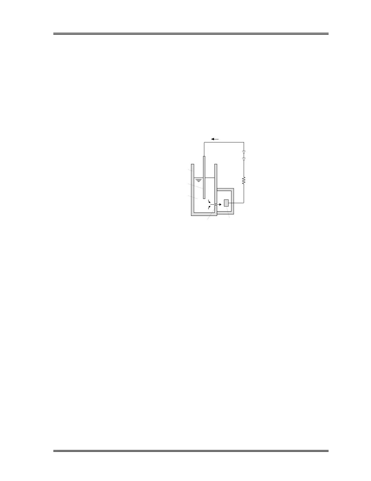
2.2 DC Detection Method
After a predetermined volume of blood is aspirated and diluted by a specific amount of
reagent or diluent, it is sent to the RBC/PLT detection chamber. In the detection
chamber, there is a small opening called an aperture. On each side of the aperture are
electrodes through which flows direct current.
When blood cells are suspended in the diluent pass through the aperture, the direct
current resistance between the electrodes changes. This resistance causes an electrical
pulse change, which is proportional to the size of the blood cell.
Data collected on the size of these pulses can be used to draw a particle size distribution
curve, which reflects the size of the blood cells. Various types of analysis data can then
be obtained from these particle size distribution curves.
Detection chamber
External electrode (+)
Electrolyte solution
Aperture Internal electro
DC s
Resis
Derect current
Figure 10-3: DC Measurement System
2.3 SLS-Hemoglobin
Conventionally, the most common automated method of measuring hemoglobin is the
cyanmethemoglobin method. This method has both advantages and disadvantages when
used with a fully automated instrument such as the SF-3000.
The cyanmethemoglobin method was recommended as the international standard in 1966
by the ICSH (International Committee for Standardization in Haematology). However,
because the speed of hemoglobin transformation is slow, this method was deemed
inappropriate for the quick processing of multiple samples by automation. Also, using
cyanide, a toxic substance, requires special disposal of the waste fluid, making the
method even less desirable.
The SLS-hemoglobin method makes use of the best parts of two methods: the
oxyhemoglobin method and the cyanmethemoglobin method.
The SLS-hemoglobin method, as with the oxyhemoglobin method, is considered to be
appropriate for automation because the transformation of blood hemoglobin is fast and
non toxic substances are used.
Further, since methemoglobin can be analyzed, control samples such as blood containing
methemoglobin can also be accurately analyzed.
In the SLS-Hg method, surfactants lyze the red blood cell membrane releasing
hemoglobin. The globin group of the hemoglobin molecule is altered by the hydrophilic
alkyl group of Sodium Lauryl Sulfate. This induces the conversion of hemoglobin from
the ferrous (Fe
+2
) to the ferric (Fe
+3
) state forming methemoglobin which combines with
Sodium Lauryl Sulfate to become SLS-Hb hemichrome molecule.