1 Use the na
vigaon toolbar to review the series and select an image to be used for calibraon.
NOTE You can change the calibraon image at any me by clicking Change in the control panel
and selecng a dierent image.
2 Click the Calibraon task.
3 Click Sphere in the Select calibraon method list.
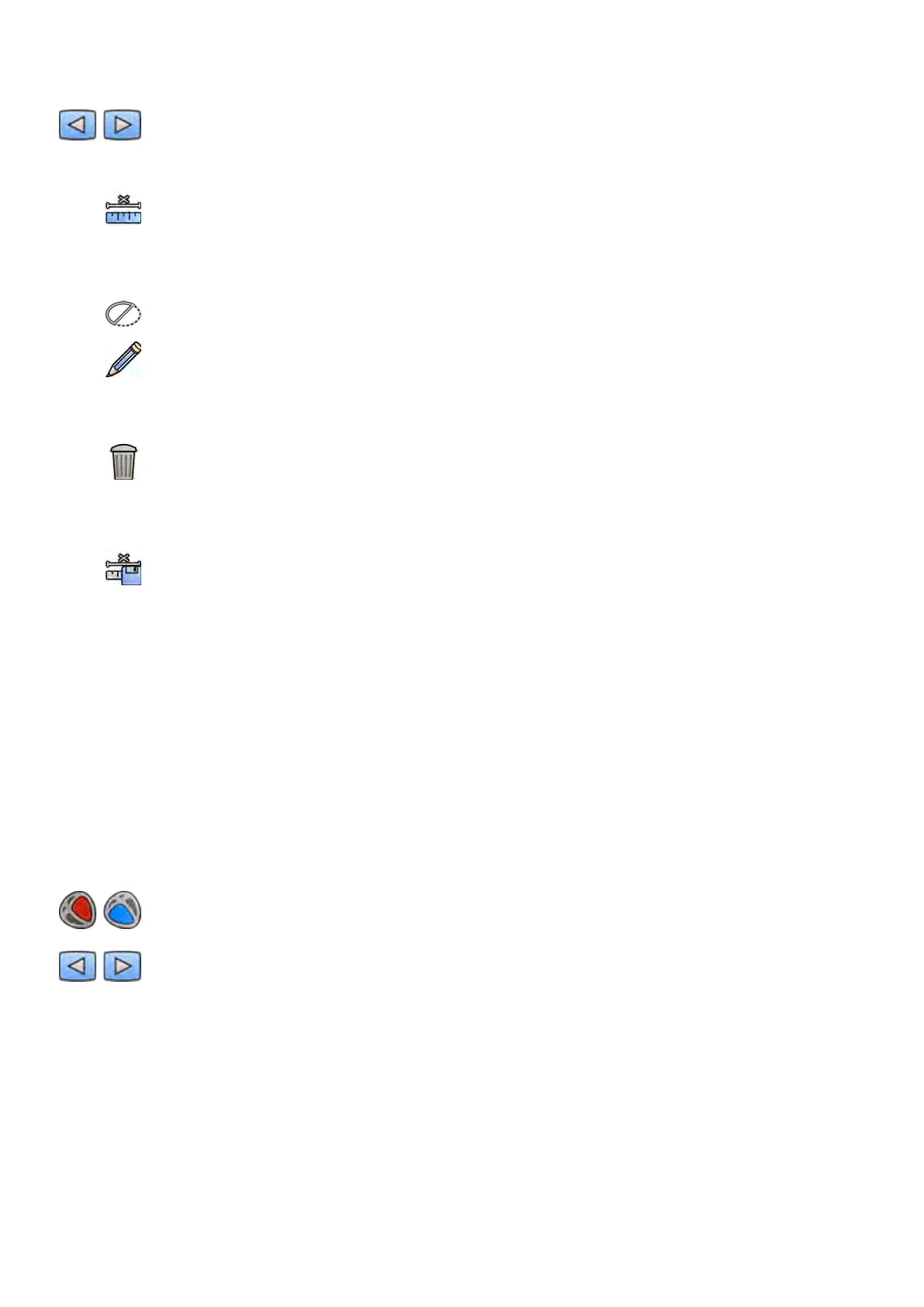
4 Click a sphere in the image to idenfy it.
5 To hide or show the sphere contour, select or clear Hide in the control panel.
6 To edit the sphere, click Edit in the control panel, and do any of the following:
• To move the sphere, drag the center of the sphere to a new posion.
• To change the diameter of the sphere, drag the circumference of the sphere.
7 If you are using Biplane LVA/RVA: Mark the sphere in both the frontal image and the lateral image.
8 You can delete the calibraon at any me and start over by clicking Delete in the task panel.
9 When the sphere is dened, select the diameter in the list in the control panel.
If the desired diameter is not available, you can type it directly in the box.
10 To accept the calibraon factor, click Accept and Connue in the control panel.
10.6.4 End Diastole (ED) Task
You use the End Diastole task to select the ED image from the series and to dene a contour on the
image.
When dening a contour in LVA, you can use either a semi-automac method or a manual method.
When dening a contour in RVA, you can only use the manual method.
Selecng the ED Image
Before you dene the ED contour, you must select a suitable image that shows the ED posion.
If the ECG is available, it is displayed with the series to assist you with idenfying the ED posion.
1 Click End Diastole in the tasks panel.
2 Use the navigaon toolbar to review the series and select an image that shows the ED posion.
Dening the ED Con
tour Semi-Automacally in LVA
To dene a contour semi-automacally in LVA, you place three key points on the selected image.
2D Quant
ave Analysis (Opon) LVA / RVA
Azurion Release 1.2 Ins
trucons for Use 171 Philips Healthcare 4522 203 52421