Using the wedges reduces the r
adiaon intensity in a user-dened area and improves the image
quality. The wedges also reduce the dose area product and the sta dose.
The amount of radiaon that is reduced by the wedges depends, for example, on the amount of the
image coverage by the wedges.
Source-to-Image Distance
According to the inverse square law, beam intensity increases proporonally with the square of the
distance.
When the source-to-image distance is increased by a factor x, the system increases the skin dose by a
factor x
2
to maintain the requested detector dose.
Hence, the source-to-image distance should be kept to a minimum (for a given source skin distance), so
the requested detector dose is reached with as low as possible skin dose. This implies that the source-
to-image distance should be reduced so that the distance between the paent and the detector is as
small as possible.
Table Height
The table height at a constant source-to-image distance does not inuence the reference air kerma
(rate), and the indicated air kerma (rate) value, as these are only applicable at the paent entrance
reference point.
It does however, inuence the paent skin dose through the inverse square law. For more informaon
on the inverse square law, see Source‐to‐Image Distance (page 293).
To minimize the skin dose (rate), the X-ray source must be as far from the skin as possible.
Inuence of Oblique Projecons
Due to the absorpon of radiaon in human ssue, the X-ray eld strength is reduced by a factor 2,
approximately every 3 cm.
For example, if the paent thickness is 27 cm, the X-ray beam loses intensity within the body by a factor
of 512 (2
(27/3)
). This shows that a thicker paent requires a larger entrance dose than a thin paent, to
obtain the same detector dose.
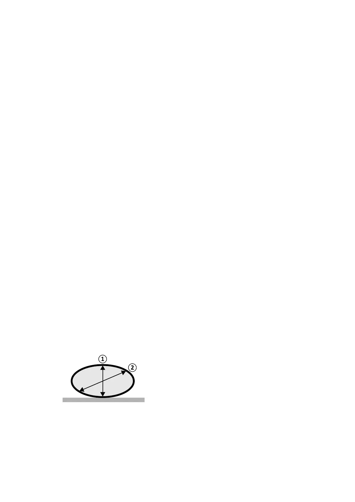
The same applies to oblique projecons of the X-ray beam since an oblique view generally increases the
perceived paent thickness. This can be seen in the gure below where distance 2 (oblique) is
considerably larger than distance 1.
Figure 132 P
aent thickness
The following example shows that the resulng air kerma is larger for a 30 cm PMMA than for a 20 cm
PMMA paent thickness, when measured at the same system sengs for three typical exposure
procedures.
Technical In
formaon System Sengs Inuencing the Radiaon Dose
Azurion Release 1.2 Ins
trucons for Use 293 Philips Healthcare 4522 203 52421